Dental Implants • 9 min read
Dental Implants in Kanpur: How Specialist Implantology Restores Missing Teeth
From the first 3D scan to the final custom ceramic crown — a complete, stage-by-stage look at modern implant dentistry and what makes it last.
A single missing molar can change the way an entire mouth works. Food starts getting stuck in the gap, the neighbouring teeth lean into the void, the opposing tooth drifts downwards, and the jawbone beneath the gap begins to shrink — quietly, from the very first month. For years, the standard answers were a bridge or a removable partial denture. Today, dental implants in Kanpur offer something fundamentally different: a titanium root placed directly into the jawbone that replaces the missing tooth at its root, not just at its crown. This guide walks through the entire journey — diagnosis, digital planning, the surgical phase, osseointegration, the custom prosthetic, and lifelong maintenance — so you know exactly what specialist implantology involves and why it remains the most durable tooth-replacement option available today.
Why One Missing Tooth Is a Bigger Deal Than It Looks
Most adults in Kanpur who lose a tooth do not lose it because of a dramatic accident. They lose it because of untreated decay, a cracked root, severe periodontal disease, or an extraction that was never followed by a replacement. The gap itself is cosmetic. The consequences are not.
The most important consequence is alveolar bone resorption. The jawbone that once held the tooth root depends on that root for stimulation. When the tooth is removed, the bone begins to resorb — a process that is fastest in the first twelve to twenty-four months and can remove a meaningful portion of ridge width and height in that window. Resorbed bone is harder to work with, may require grafting, and changes the shape of the face over time, giving the cheeks and lips a sunken appearance. A bridge or a partial denture replaces the visible tooth but does nothing to stop this.
- ✓ Tooth drift and tipping: adjacent teeth rotate and lean into the gap, widening the space and making cleaning harder.
- ✓ Over-eruption: the opposing tooth grows downwards into the void, altering the bite and stressing the jaw joint.
- ✓ Occlusal overload: a shifted bite concentrates chewing forces on a few teeth, accelerating wear, fractures and gum recession.
- ✓ Food trapping and decay: retained food debris at the gap is a common trigger for new cavities and gum inflammation.
Implants vs Bridges vs Dentures: An Honest Comparison
When patients visit an implant dentist in Kanpur for a single missing tooth, three options are usually discussed. All of them replace the missing tooth. They differ profoundly in how they do it, how long they last, and what they ask of the surrounding teeth. A conventional three-unit bridge, for example, requires the two healthy teeth on either side of the gap to be cut down substantially to accept abutment crowns — a preparation that is irreversible. An implant avoids that entirely because it is supported by bone, not by teeth.
Typical reported service life by replacement type
Conservative published ranges (midpoint shown). Individual outcomes vary with bone quality, hygiene, occlusion and aftercare.
Implant-supported crowns are the only option shown that also preserves alveolar bone.
| Criteria | Dental Implant | Conventional Bridge | Removable Partial Denture |
|---|---|---|---|
| Support | Directly integrated with jawbone | Cemented/crowned onto adjacent teeth | Gum tissue and clasps on remaining teeth |
| Effect on natural teeth | None — adjacent teeth untouched | Irreversible reduction of healthy enamel | Minimal, but clasps may wear enamel over time |
| Bone preservation | Functional load maintains ridge density | No stimulation; ridge resorbs beneath pontic | No stimulation; resorption continues |
| Typical lifespan | 20–30+ years; implant body often retained for life | 5–15 years before replacement | 5–10 years; relining and rebasing needed |
| Maintenance | Brushing, interdental cleaning, 6-month reviews | Superfloss under pontic, higher decay risk at margins | Daily removal and cleaning; night-time removal |
| Comfort / function | Feels like a natural tooth; normal chewing force | Feels natural; slight difference in feedback | Bulk, movement and clicking often reported |
| Best suited for | Most single-tooth cases with adequate bone | Cases where implants are not viable | Multiple missing teeth, temporary or budget-limited solutions |
Bridges remain a perfectly reasonable option in some situations — when bone volume is insufficient without grafting, when a patient prefers a shorter treatment, or when adjacent teeth already need crowns. But for a lone missing tooth with healthy neighbours, implantology is the more conservative long-term choice because it protects the very teeth a bridge would sacrifice.
The Consultation, Digital Imaging and Treatment Planning Stage
Nothing in implant dentistry is more important than what happens before the drill touches the mouth. A thorough assessment usually takes 45 to 60 minutes and covers three dimensions: biological, mechanical and aesthetic.
Biological assessment means evaluating gum health, the condition of adjacent teeth, plaque control, and the general medical history — particularly uncontrolled diabetes, hypertension, bleeding disorders, osteoporosis medications, and smoking or tobacco chewing habits. Mechanical assessment involves occlusion analysis: how the teeth actually meet, whether there is heavy grinding or clenching, and how much load the future implant will carry. Aesthetic assessment maps the gum line, papillae, lip line and smile zone, so the final crown emerges from the gum exactly where a natural tooth would.
Digital imaging completes the picture. A periapical radiograph shows fine root and bone detail, while a low-dose CBCT (cone-beam computed tomography) scan provides a true 3D volume of the jaw — revealing bone width, height, density, the position of the inferior alveolar nerve in the lower jaw and the maxillary sinus in the upper jaw, and the exact location of neighbouring roots. Intraoral scanning then captures a digital impression without the gagging, gagged-altered gag reflex and tray discomfort many patients associate with moulding. Combined with guided-surgery software, this data allows the surgeon to place the implant on a screen before entering the mouth.
The implant journey at a glance
Click any column header to sort. Timelines are typical and vary by case.
Titanium Implants, Bone Grafting and the Surgical Phase
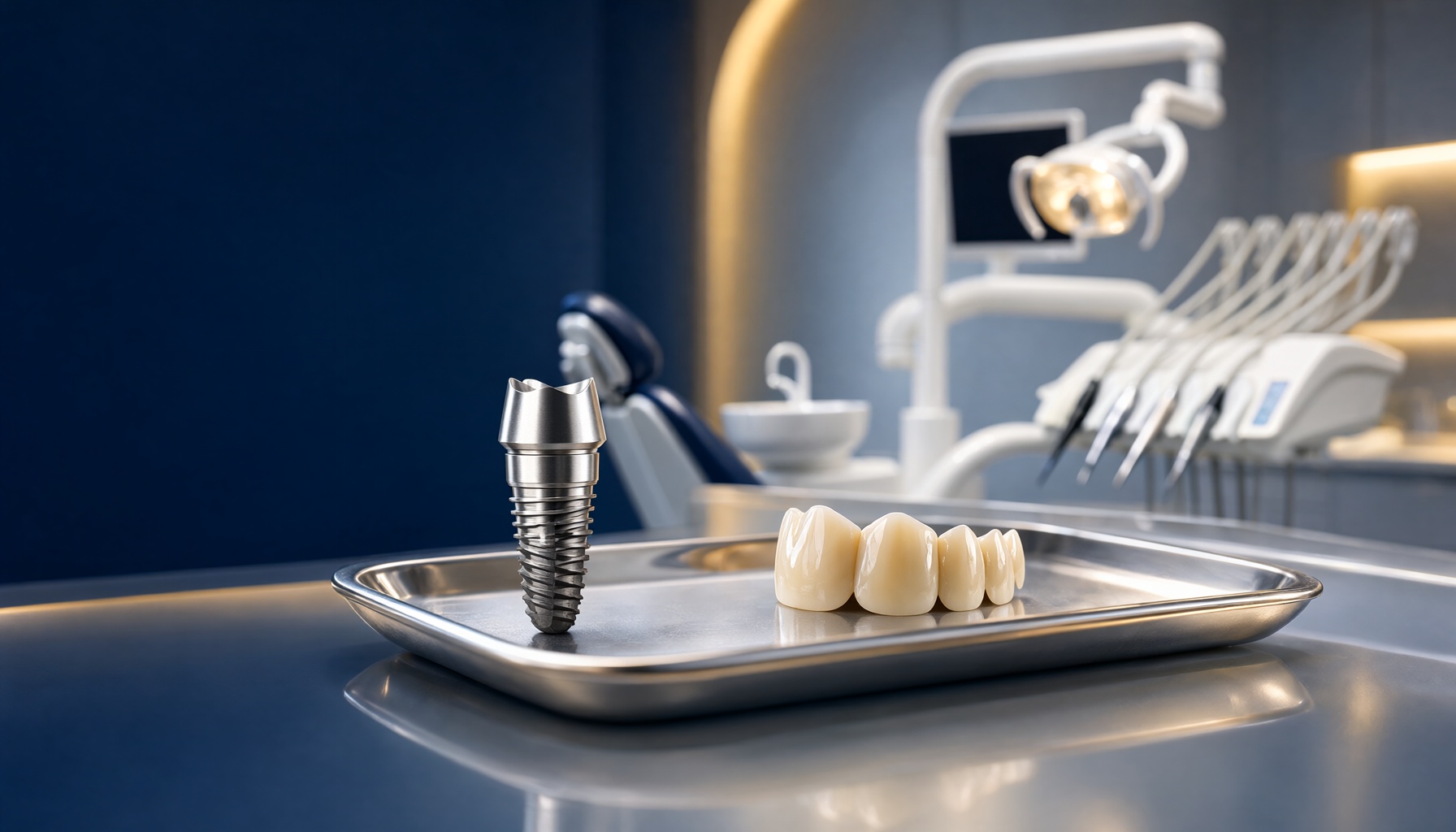
A standard implant is a precision-machined, screw-shaped post made from titanium or a titanium–zirconium alloy. Its surface is treated — by acid etching, sandblasting, anodic oxidation or laser micro-texturing — to create a micro-rough architecture that encourages bone cells to attach rapidly. The implant carries three principal parts: the fixture (the part inside the bone), the abutment (the connector above the gumline), and the crown (the visible, custom-fabricated tooth).
Plate-like or screw-shaped implants of different diameters and lengths are selected according to the space available. Wider implants (4.5–6 mm in the posterior jaw) distribute occlusal load more favourably, while shorter narrow-diameter implants are used where the inter-root space is tight or bone height is limited. Implant number, angulation and emergence profile are all decided in the planning stage, not at the chairside.
Bone grafting: why augmentation is sometimes necessary
Schematic illustration of decision thresholds commonly used in implant planning, not patient-specific measurement.
If measured bone falls short of the ideal envelope, augmentation is performed before or at implant placement.
Bone grafting fills a deficit so that an implant can be placed in the correct prosthetic position. Autogenous bone (harvested from the patient) remains the gold standard because it contains living osteogenic cells, but it requires a second surgical site and is limited in volume. Xenografts, allografts and synthetic materials are therefore widely used, often alone or in composite form. Guided bone regeneration (GBR) uses a membrane — collagen, resorbable or non-resorbable — to exclude soft tissue and preserve a space for bone to grow in. Socket preservation, placing graft material into the extraction socket at the time of the extraction, is one of the most effective ways to prevent ridge loss altogether.
The surgical phase itself is usually performed under local anaesthesia. A small pilot drill, progressively larger drills and finally the implant itself are advanced along the planned trajectory, typically at 1–2 mm above the adjacent gum crest. Once torque reaches the insertion torque value specified by the system, primary mechanical stability is achieved. In suitable cases, a healing cap or temporary crown can be attached in the same visit — a protocol known as immediate loading.
Osseointegration, the Healing Timeline and the Custom Crown
Osseointegration is the biological process at the heart of implantology: direct structural and functional connection between living bone and the titanium implant surface, with no intervening fibrous tissue. It was first described by the Swedish surgeon Per-Ingvar Brånemark in the 1960s and remains the single most important determinant of long-term implant success.
Healing phases after implant placement (illustrative timeline)
Time ranges vary with bone quality, graft volume, smoking status and healing capacity.
The crown is fabricated only after stability and soft-tissue maturation are confirmed.
Weeks 0–2 — Inflammation and clot phase
The blood clot forms around the titanium surface. Cells of the immune system clear debris while the gum flap or cover screw is repositioned and sutured. Most swelling resolves during this window.
Weeks 2–6 — Proliferation and woven bone
New blood vessels grow in, and osteoblasts lay down immature woven bone directly against the implant threads. The primary mechanical stability gradually converts into biological stability.
Weeks 6–12 — Early maturation
Woven bone remodels into lamellar bone. If grafting was performed, its mineralisation continues in parallel. A stability check — often resonance frequency or percussion — may be carried out around this point.
Months 3–6 — Secondary stability and soft-tissue shaping
The bone–implant interface achieves secondary stability. In posterior sites and grafted cases this phase commonly extends to six months, sometimes longer.
Months 4–6 — Final scan, fabrication and fit
A scan or impression captures the healed soft tissue, a custom abutment is selected, and the zirconia or lithium-disilicate crown is milled. The restoration is tried in, shade-matched and cemented or screw-retained.
The prosthetic phase is where implantology becomes visible artistry. High-strength zirconia, lithium-disilicate and porcelain-fused-to-metal remain the dominant crown materials; each is layered or monolithic, characterised from a shade tab under a standardised light source, and adjusted so that emergence profile and gum contour mimic the adjacent teeth. Screw-retained designs, where a channel is prepared through the crown and the abutment is fastened from inside, allow the restoration to be removed non-destructively — a significant long-term maintenance advantage over cement.
“An implant is the only tooth replacement that is handed back to the jawbone itself. That is why it is the only one that can stop the bone loss other options accelerate.”
Aftercare, Long-Term Maintenance and the Causes of Implant Failure
The implant is placed in month one; the relationship is built for decades. A structured maintenance schedule is the single most effective predictor of implant survival, and it is something a responsible implant dentist in Kanpur will always schedule before the final crown is delivered.
Reported implant survival by years in function
Longitudinal survival figures compiled from published clinical literature and systematic reviews.
Survival declines gradually; catastrophic loss is uncommon and usually has a identifiable cause.
Day-to-day care mirrors care for natural teeth: brushing twice daily, with an electric brush and a fluoride or implant-specific paste; interdental brushes, floss threaders or water flossers passed under and around the crown; and avoidance of biting hard objects such as ice, nuts and hard candy on the implant crown. Professional review every six months allows the dentist to measure pocket depths, assess bleeding on probing, check occlusion and clean around the crown margin. Where a patient is a grinder, a night guard is a routine and inexpensive protective measure.
Failure is uncommon but it does occur, and understanding it matters. Peri-implantitis — an inflammatory destruction of bone around a functioning implant, analogous to periodontitis — is the most frequent late cause of loss, and is driven primarily by biofilm. Biological failure in the early phase presents as early osseointegration failure, where insufficient initial stability, contaminated site, inadequate vascularisation or heavy early loading prevents bone formation. Mechanical overload from bruxism, a heavy bite or an unfavourable prosthetic design causes screw loosening, abutment fracture, or prosthetic screw fracture. Systemic and behavioural factors — smoking, uncontrolled diabetes, poor plaque control, osteoporosis, and non-compliance with follow-up — consistently appear in the literature as risk multipliers. Crucially, many early failures are salvageable if identified promptly, which is exactly why the maintenance schedule is not optional.
| When | Sign | Why it matters |
|---|---|---|
| First week post-surgery | Increasing pain, swelling, fever, pus discharge | Possible early infection; may need debridement or early intervention |
| First 3 months | Implant feels loose or the crown is tender on biting | Suggestive of incomplete osseointegration or overload |
| Years 1–5 | Bleeding or swelling of gum tissue around the crown | Early peri-implant mucositis — treatable and reversible at this stage |
| Any year | Metal visibility, persistent bad taste, recession of the gum margin | Progressive bone loss; a radiograph is required promptly |
Key Takeaways
A missing tooth is a moving target for the rest of the mouth, and the bone loss that follows begins almost immediately. When you compare dental implants in Kanpur against bridges and partial dentures, the distinction becomes clear: an implant is the only option that restores both the crown and the root function, preserves the jawbone, and leaves the neighbouring teeth completely untouched. Reported long-term survival sits above 90% across most published studies, with a realistic expected service life measured in decades rather than years.
The outcome, however, is determined less by the implant brand than by the team and the plan behind it: accurate 3D diagnosis, correct 3D positioning, biological respect for bone, and a patient who commits to maintenance. For anyone in Kanpur weighing their options, the right first step is not a product decision — it is a comprehensive implant consultation with a qualified implant dentist in Kanpur, ideally one who will show you your own scan, explain the bone you actually have, and be candid about whether an implant is genuinely the best choice for your mouth.
Ready to Restore Your Smile?
Schedule a detailed implant consultation at The Crown Multispeciality Dental Clinic & Implant Center, Kanpur. We will take a digital scan, assess your bone and gum health, and walk you through the complete treatment sequence before you make any decision — no pressure, no guesswork.
Book Your Implant ConsultationExplore more about our dental implant services and general preventive oral care.
Related Articles
Tooth Pain When Biting Down in Kanpur: 4 Common Causes
General Dentistry · 9 min read Tooth Hurts When You Bite Down: Four Common Causes Explained by Kanpur…
Cracked Tooth Repair in Kanpur: Save It or Remove It?
Restorative Dentistry · Kidwai Nagar, Kanpur Cracked Tooth Repair in Kanpur: How a Dentist Decides Between Saving a…
How Do Braces Work in Kanpur? The Biology of Tooth Movement
Orthodontics • 14 min read How Do Braces Work in Kanpur? The biology behind tooth movement — what…