Dental Implants • 10 min read
Dental Implants in Kanpur: How Specialist Implantology Restores Missing Teeth
A clinically proven, biologically guided route back to a natural-looking, fully functional smile — planned digitally, placed precisely and maintained for decades.
Losing a single tooth is rarely just a cosmetic event. The moment a tooth is extracted, the jawbone beneath it begins a silent, irreversible process of resorption — and the two teeth on either side start to tip and drift. For thousands of patients across Kanpur, the answer today is dental implants in Kanpur: a titanium root that is placed into the bone, fuses with it, and carries a custom-made ceramic crown that looks and functions like the tooth you lost. This guide walks through the entire journey — from the first consultation to long-term maintenance — and explains why choosing a qualified implant dentist in Kanpur is the single most important decision in the process.
Chapter 1: Why Single Tooth Loss Is a Growing Concern Among Adults in Kanpur
India’s population is ageing, and with it the burden of edentulism and partial tooth loss. Sugar consumption, tobacco use, untreated periodontal disease and diabetes — all highly prevalent in the region — mean more adults in their 40s, 50s and 60s are now presenting with missing teeth than ever before. National oral health data consistently shows periodontal disease affecting roughly half of all adults, and it is the leading cause of tooth loss in people under 45.
What makes single tooth loss a genuine clinical concern is not the missing tooth itself but the biological cascade that follows. A dental implant in Kanpur addresses every one of those consequences, which is why implantology has become the backbone of modern restorative dentistry in the city.
- ✓ Bone resorption: without a root to load the socket, the alveolar ridge loses a large share of its height and width within the first year.
- ✓ Tipping and drifting of adjacent teeth: neighbours lean into the gap, ruining contacts, bite forces and the smile line.
- ✓ Reduced chewing efficiency and jaw health: one-sided chewing can strain the temporomandibular joint (TMJ) and alter digestion.
- ✓ Speech, nutrition and confidence: gaps in the front teeth alter certain sounds; limitations in chewing restrict food choices.
What Actually Happens to the Jawbone After Extraction?
The ridge under a lost tooth remodels rapidly. Clinical data shows the greatest width reduction occurs in the first six to twelve months, after which resorption continues more slowly but never fully stops. This is precisely the biology that makes an implant unique: once the titanium fixture is integrated and loaded by a crown, the chewing forces transmitted through it maintain — and can partially rebuild — the bone.
Chart 1 — Alveolar ridge height loss over time after tooth loss
Illustrative average reduction in vertical ridge height at the extraction site.
Chapter 2: Implants, Bridges and Dentures Compared on Longevity, Maintenance and Bone Health
Patients usually arrive with three options in mind. A resin-bonded bridge is the most conservative, requiring minimal preparation but a shorter expected service life. A conventional tooth-supported bridge is durable but requires healthy teeth on both sides of the gap to be filed down as abutments. A removable denture or partial denture is the least invasive and the least stable, needing periodic relining and often affecting the bone negatively. A standalone dental implant replaces only the missing tooth, protects the neighbours and preserves bone load.
Table 1 — Head-to-head comparison of tooth replacement options
Typical reported outcomes for a single missing tooth in healthy adult patients.
Long-term survival studies of endosseous implants — from the original Brånemark work through to modern systematic reviews — consistently report single-tooth implant survival in the high 90s over a decade, with zirconia or porcelain-fused-to-metal crowns wearing out slightly before the fixture itself. A bridge, by contrast, is usually limited by the weakest abutment tooth, and every abutment preparation is a permanent commitment.
Chart 2 — Expected service life by replacement option
Average years in service before replacement or major repair is typically required.
Chapter 3: The Consultation, Digital Imaging and Treatment Planning Stage
Implantology is a diagnosis-first specialty. A specialist implant dentist in Kanpur will rarely book surgery on the first visit. Instead, the process begins with a structured assessment that typically includes a medical and dental history, a full periodontal charting of the remaining teeth, an examination of the bite, photographs of the smile, and a discussion of the patient’s expectations.
Why 3D imaging changed everything
A conventional periapical X-ray shows a two-dimensional slice and cannot tell you what lies buccally, lingually or behind the sinus. CBCT (cone-beam computed tomography) gives a true three-dimensional volume in a fraction of a second of exposure, allowing the surgeon to measure available bone height and width, identify critical structures such as the inferior alveolar nerve and the maxillary sinus floor, and confirm inter-root distances. With that data loaded into planning software, the entire implant position, diameter and angle can be simulated on screen, and a surgical guide can be 3D-printed to transfer that virtual plan to the patient’s mouth with sub-millimetre accuracy.
Guided surgery is not a substitute for clinical judgement — soft tissue thickness, gingival biotype and final restorative positioning still require expert planning — but it removes the guesswork from the drilling stage and dramatically reduces surgical time. Patients are also shown a digital preview of the final tooth before treatment begins, which makes the decision far easier.
Chapter 4: Titanium Implants, Bone Grafting and the Surgical Phase Explained
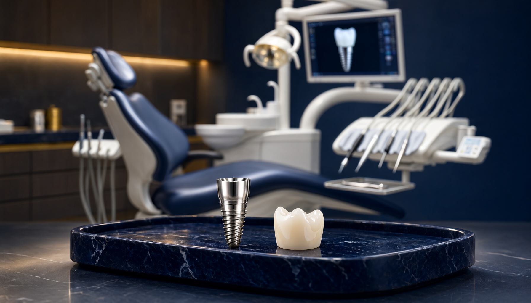
Almost all modern implants are made of commercially pure titanium or a titanium-zirconium alloy. Titanium is used because it is biocompatible and, uniquely, forms a direct chemical and structural bond with living bone — a process called osseointegration. Implant surfaces are usually treated (anodised, sandblasted, acid-etched or coated) to increase their surface area and accelerate bone growth. The vast majority of implants placed today are endosseous fixtures: threaded, screw-shaped roots available in lengths of roughly 6–16 mm and diameters of 3–5 mm, with the shape and thread depth chosen for the density of the local bone.
When bone grafting is needed
If the CBCT shows insufficient bone to place an implant in a prosthetically correct position, the site is augmented before or at the time of implant placement. Graft materials fall into four families: autograft (bone taken from the patient’s own jaw — the gold standard for osteogenic potential), allograft (donor bone, highly osteoconductive), xenograft (bovine or porcine mineral) and alloplast (synthetic materials). Sinus lift procedures augment the bone floor of the maxillary sinus for posterior upper teeth; ridge augmentation widens a deficient ridge. Healing after grafting typically takes four to six months before the implant is placed.
The surgical appointment itself is usually performed under local anaesthesia. After a small incision, the specialist prepares a precise osteotomy with sequential drills, places the fixture at the planned depth and angle, and closes the site. Guided techniques allow a flapless approach, meaning no stitches and very little postoperative swelling.
Chapter 5: Osseointegration, the Healing Timeline and the Custom Prosthetic Crown
Osseointegration begins within days of placement but matures over months. Blood clot first fills the gap, then osteoblasts deposit woven bone on the implant surface, which is progressively remodelled into mature lamellar bone. The lower jaw, being denser and more rigid, typically osseointegrates in three to four months; the softer maxillary bone usually needs four to six months. In selected cases — with good primary stability and favourable bone — immediate loading or early loading protocols can shorten this, but they are only appropriate for carefully selected cases.
Chart 3 — The implant treatment timeline
Progression of biological integration and prosthetic stages over the first six months.
Once integration is confirmed, the restorative phase begins. An impression or digital scan captures the implant position, and the laboratory fabricates a screw-retained or cement-retained crown. The most common materials today are zirconia and lithium disilicate — both metal-free, highly translucent and biocompatible — often layered over a porcelain-fused-to-metal coping where extra strength is needed. The crown is matched not just to the shade of the neighbouring teeth but to their translucency, fluorescence and surface texture, so that in daylight the implant crown is genuinely indistinguishable. In aesthetic front-tooth cases, a small amount of pink ceramic or connective-tissue grafting may be used to recreate a natural gum contour.
“The implant is not the tooth. The implant is the foundation. The tooth is whatever you and your prosthetist decide to build on it — and it should be built to protect the bone for the rest of your life.”
Chapter 6: Aftercare, Long-Term Maintenance and What Causes Implant Failure
An implant does not fail suddenly; it is usually lost to a slow inflammatory process. Peri-implantitis is the destructive, bone-resorbing infection of the tissues around an implant, analogous to periodontitis. It is initiated by plaque biofilm and accelerated by smoking, poorly controlled diabetes, a history of periodontitis, bruxism and the absence of professional maintenance. Peri-implant mucositis — inflammation without bone loss — is far more common and is fully reversible when caught early.
Long-term care is straightforward. Clean the implant with a soft brush and curved interdental brushes or a water flosser, be careful not to let plaque sit around the gum margin, treat clenching or grinding with a night guard, control diabetes and stop smoking, and attend scheduled maintenance visits — typically every three to six months. At these visits the implant is measured for pocket depths, mobility and bleeding, radiographs are taken periodically to compare bone levels, and the crown is polished and checked for occlusion.
Table 2 — Risk factors for implant complications and how to manage them
Understanding these factors is the foundation of predictable implantology.
Interestingly, the prosthetic crown is usually the first thing to need attention, not the implant. Chips, wear or cement remnants can be repaired or replaced relatively easily — which is one of the great practical advantages of a screw-retained, retrievable design.
Why Specialist Qualification Matters
Implant surgery is a precise microsurgical discipline that sits at the intersection of oral surgery, prosthodontics, periodontology and radiology. When you search for an implant dentist in Kanpur, look for specific training credentials, the use of 3D imaging and guided surgery, an in-house or closely associated prosthodontic workflow, and a documented follow-up protocol. A multidisciplinary team — surgeon, prosthodontist, hygienist and technician — consistently achieves better outcomes than a single clinician working alone. You can explore the services offered at The Crown Multispeciality Dental Clinic & Implant Center or browse our dental implants resources for more in-depth guides.
Key Takeaways
Tooth loss is the beginning of a biological process, not an isolated problem. An implant arrests that process by restoring the load that keeps bone healthy, and among all replacement options it is the only one that protects the neighbouring teeth and the ridge simultaneously.
When comparing dental implants in Kanpur against bridges and dentures, the evidence favours implants on longevity, bone preservation and quality of life — provided the case is well planned and the patient commits to maintenance. CBCT imaging, surgical guides and a properly timed loading protocol reduce risk further, while titanium’s predictable osseointegration underpins every long-term success story.
The final takeaway is simple: the implant itself is permanent, but the outcome depends on three things — the quality of the bone, the skill of the surgeon and the discipline of the patient. Choose an experienced implant dentist, follow the aftercare plan, and a single tooth lost today can stay functionally and aesthetically intact for the rest of your life.
Missing a Tooth? Let’s Plan It Properly.
Book a consultation with our implantology team for a full clinical assessment, 3D imaging and a personalised treatment roadmap — explained clearly, without jargon.
Book Your Implant ConsultationRelated Articles
Tooth Pain When Biting Down in Kanpur: 4 Common Causes
General Dentistry · 9 min read Tooth Hurts When You Bite Down: Four Common Causes Explained by Kanpur…
Cracked Tooth Repair in Kanpur: Save It or Remove It?
Restorative Dentistry · Kidwai Nagar, Kanpur Cracked Tooth Repair in Kanpur: How a Dentist Decides Between Saving a…
How Do Braces Work in Kanpur? The Biology of Tooth Movement
Orthodontics • 14 min read How Do Braces Work in Kanpur? The biology behind tooth movement — what…